- Shopping, made easy.
- /
- Get the app!
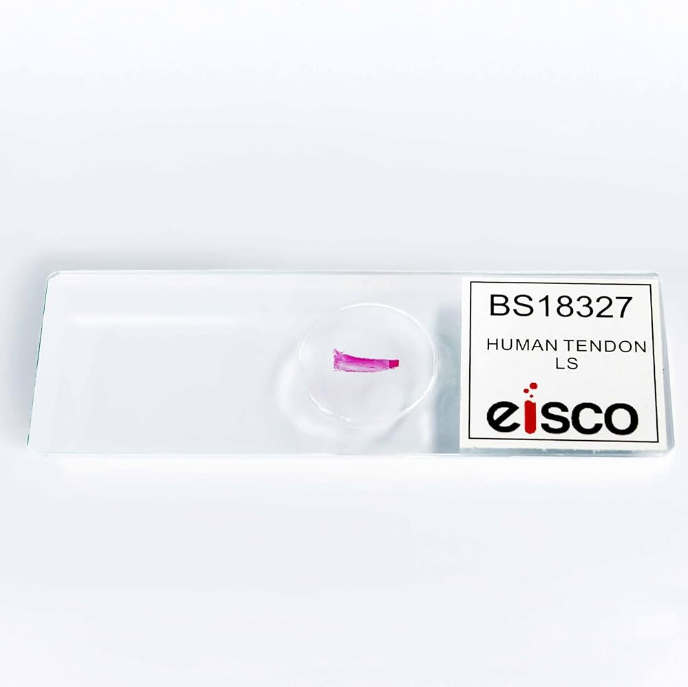
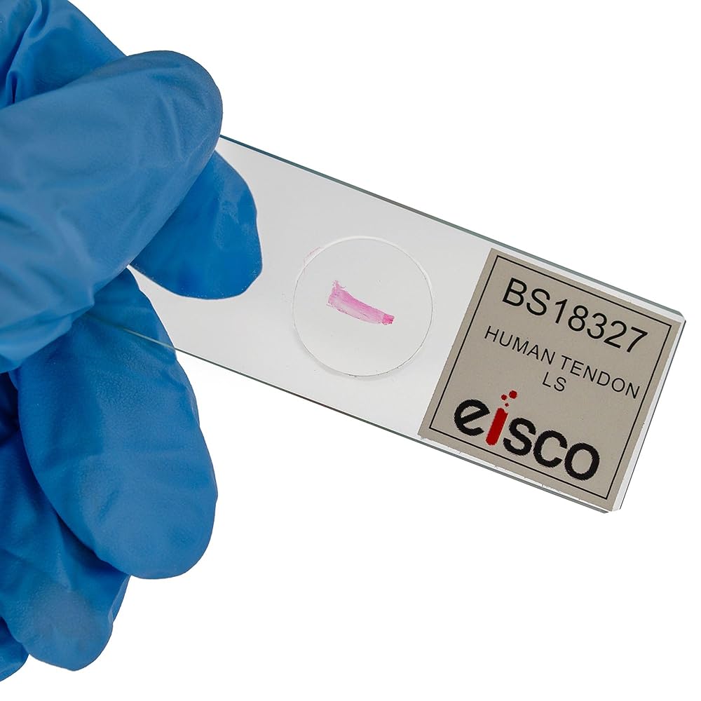
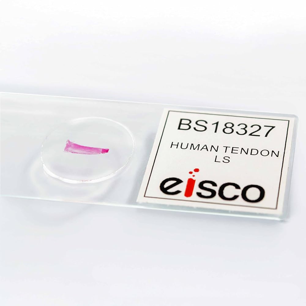
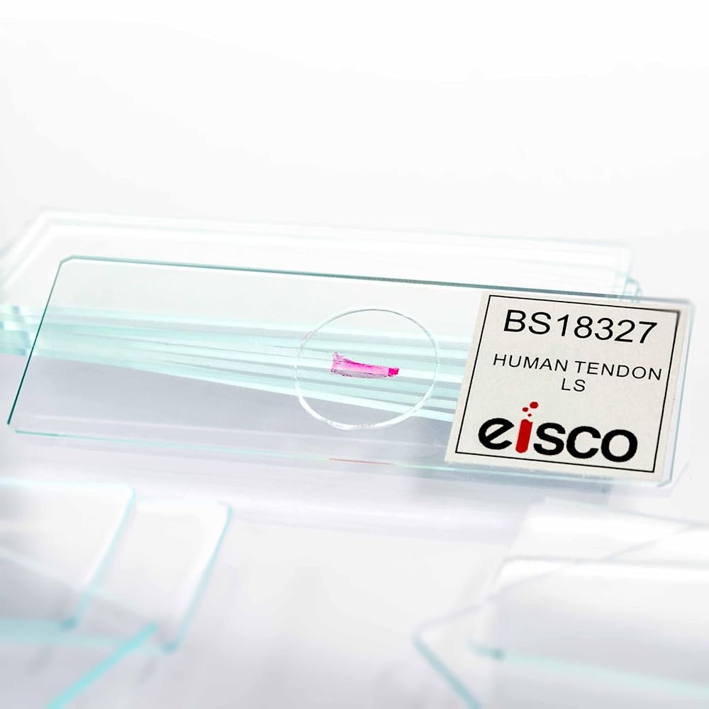
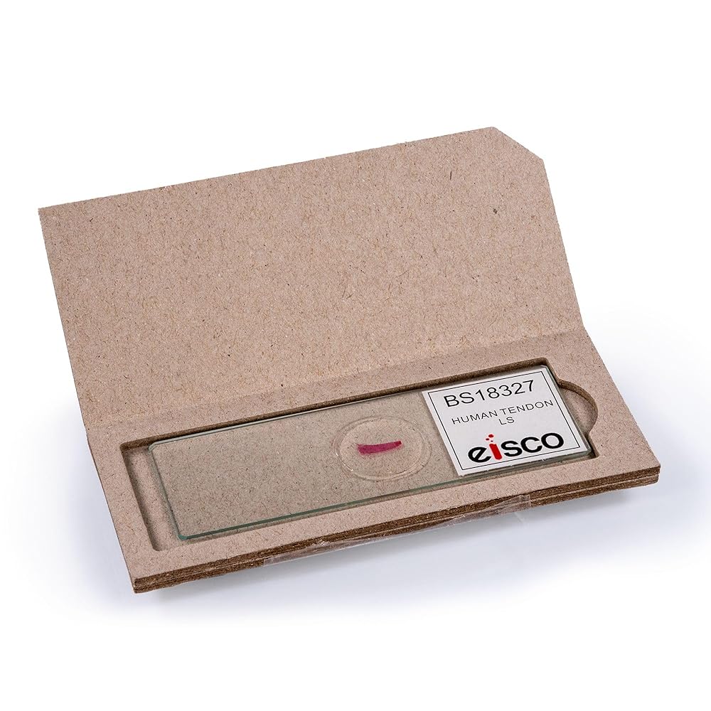
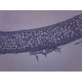
This prepared microscope slide features a longitudinal section of human tendon tissue mounted and sealed for microscopic examination. Clearly reveals the parallel alignment of collagen fibers and overall tendon structure. Supports instruction in histology, anatomy, and musculoskeletal function. Ideal for classroom demonstrations, lab-based learning, and anatomical tissue identification using standard compound microscopes.
 EISCO 10pk Spongy Bone Section (Mammal), Prepared Microscope Slides - 75 x 25mm Classroom Pack, 10 in Storage Case Anatomy, Biology & Microscopy- Eisco Labs
KWD 10
EISCO 10pk Spongy Bone Section (Mammal), Prepared Microscope Slides - 75 x 25mm Classroom Pack, 10 in Storage Case Anatomy, Biology & Microscopy- Eisco Labs
KWD 10
 10PK Yellow Elastic Cartilage - Prepared Microscope Slides - Classroom Pack, 10 Slides in Storage Case - Biology & Microscopy - Eisco Labs
KWD 6.500
10PK Yellow Elastic Cartilage - Prepared Microscope Slides - Classroom Pack, 10 Slides in Storage Case - Biology & Microscopy - Eisco Labs
KWD 6.500
 EISCO 10PK Simple Cuboidal Epithelium - Prepared Microscope Slides - Classroom Pack, 10 Slides in Storage Case - Biology & Microscopy
KWD 7.500
EISCO 10PK Simple Cuboidal Epithelium - Prepared Microscope Slides - Classroom Pack, 10 Slides in Storage Case - Biology & Microscopy
KWD 7.500
 EISCO - Ranunculus Root - Cross Section - Prepared Microscope Slide - 75 x 25mm - Biology & Microscopy
KWD 3.500
EISCO - Ranunculus Root - Cross Section - Prepared Microscope Slide - 75 x 25mm - Biology & Microscopy
KWD 3.500